

Published and medically reviewed: 20 August 2026. This page is for general education; diagnosis and an individual surgical plan require an examination.
Purpose of this guide: to explain tuberous or tubular breasts through the relationship between the breast base, lower pole, inframammary fold, areola and volume, rather than treating the issue as “small breasts” alone. Related information is available on breast augmentation, breast lift and reduction and breast asymmetry.
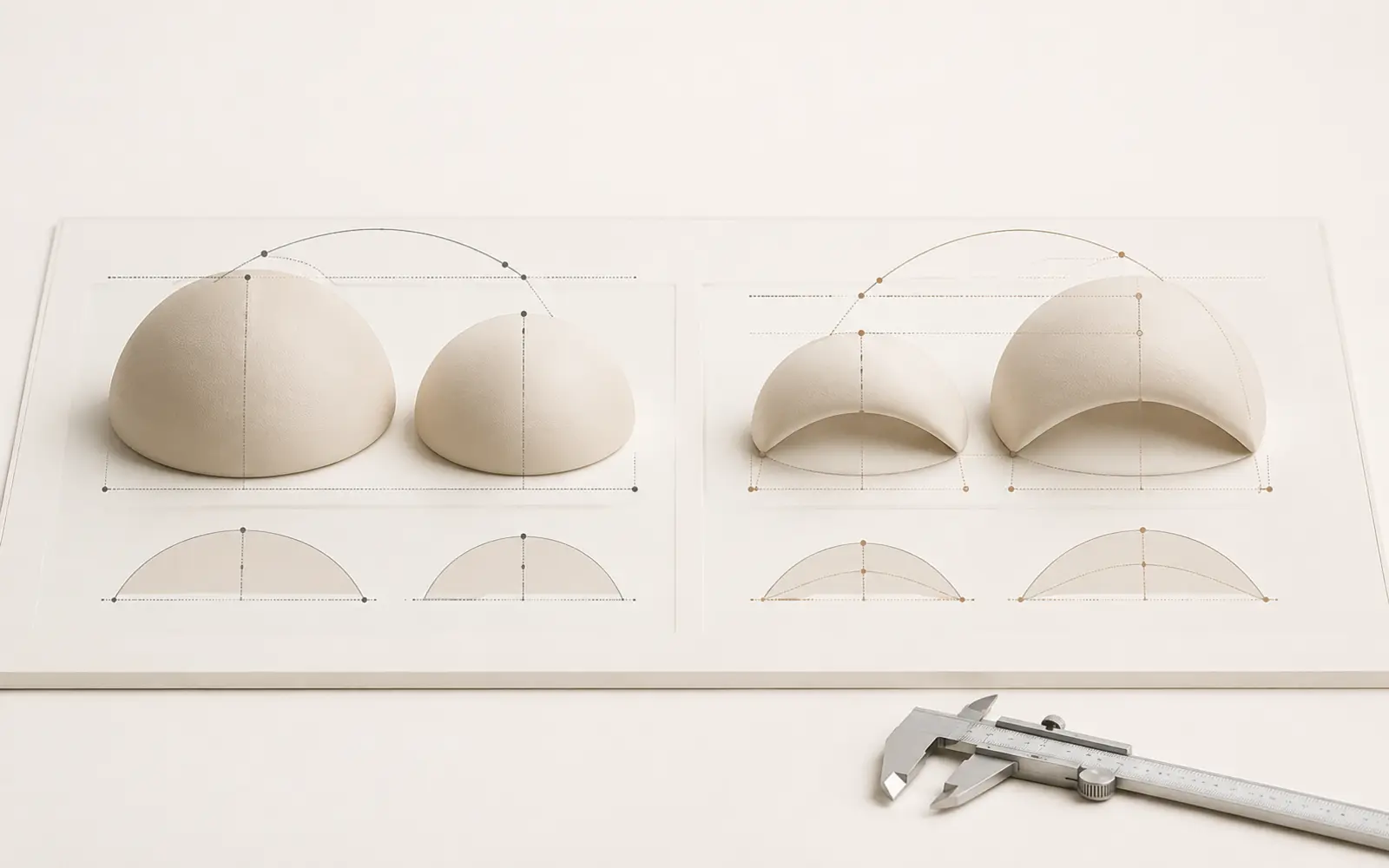
Tuberous breasts are a developmental breast-shape variation in which the base may remain constricted and the lower part of the breast may not expand as expected. The pattern can affect one or both sides, and the degree does not have to be equal. One person may have a subtle lower-pole shortage; another may also have a high fold, enlarged or prominent areolae and marked asymmetry.
“Tuberous” and “tubular” describe a spectrum rather than one fixed appearance. Constriction at the breast base may limit horizontal and vertical expansion of the lower pole. Tissue can project forward over a narrow base, the inframammary fold may sit high, and breast tissue may protrude through the areola.
The pattern usually becomes apparent as the breasts develop during puberty. It is not caused by underwear, weight control or something the patient has done. A newly appearing one-sided enlargement, lump, persistent redness, skin tethering or spontaneous discharge must nevertheless be assessed as a breast-health issue rather than assumed to be developmental.
| Pattern | Dominant feature | Main planning question |
|---|---|---|
| Simple volume asymmetry | Similar base and fold, different volume | How can volume be balanced? |
| Breast ptosis | Nipple position and loose skin dominate | Is a lift and scar pattern required? |
| General hypoplasia | Balanced base shape with low volume | Are the tissues suitable for an implant or fat grafting? |
| Tuberous pattern | Constricted base, lower-pole shortage, high fold and/or areolar change | Which structures need release and reshaping before volume is added? |
The same patient may have tuberous anatomy, volume asymmetry and ptosis together. A photograph therefore cannot establish that an implant alone is enough or that a lift is mandatory.
Each breast is assessed separately and in relation to the chest wall. Measurements include base width, nipple-to-fold distance, fold level, lower-pole flexibility, areolar diameter and prominence, skin quality, tissue thickness and chest-wall asymmetry. Previous surgery, weight change, pregnancy and breastfeeding plans, medication and nicotine exposure also affect planning.
Photographs and a remote consultation can help organise travel, but cannot show tissue tightness, fold mobility or a palpable finding. Ultrasound, mammography or another test may be requested according to age, history and examination.
Tight tissue that limits lower-pole expansion can be carefully released or rearranged. The aim is not merely to enlarge the breast, but to allow the base and lower pole to take on a more balanced shape.
A high fold may be lowered or balanced between sides according to the planned lower-pole length. Precise positioning matters because an inadequately controlled fold can contribute to a double-contour appearance.
An implant may be used when the tissues and volume goal are suitable. Selection involves base width, tissue coverage, profile and side-to-side differences—not just implant volume. Breast implants are not lifetime devices and can require future monitoring or surgery.
Fat taken from a suitable donor area can support lower-pole expansion or smooth contour transitions. Retention varies, and one session cannot promise exact correction of a marked constriction.
A widened or prominent areola may be adjusted through a periareolar approach. A lift may be added when nipple position or loose skin requires it. Tension, scar widening and recurrent areolar prominence are part of informed discussion.
Tuberous does not always mean small. When the breast is large and heavy, reduction, base reshaping and lifting may be planned together.
Marked constriction, a limited skin envelope or major asymmetry may be treated in stages, using tissue expansion or fat grafting first and an implant or further reshaping later.
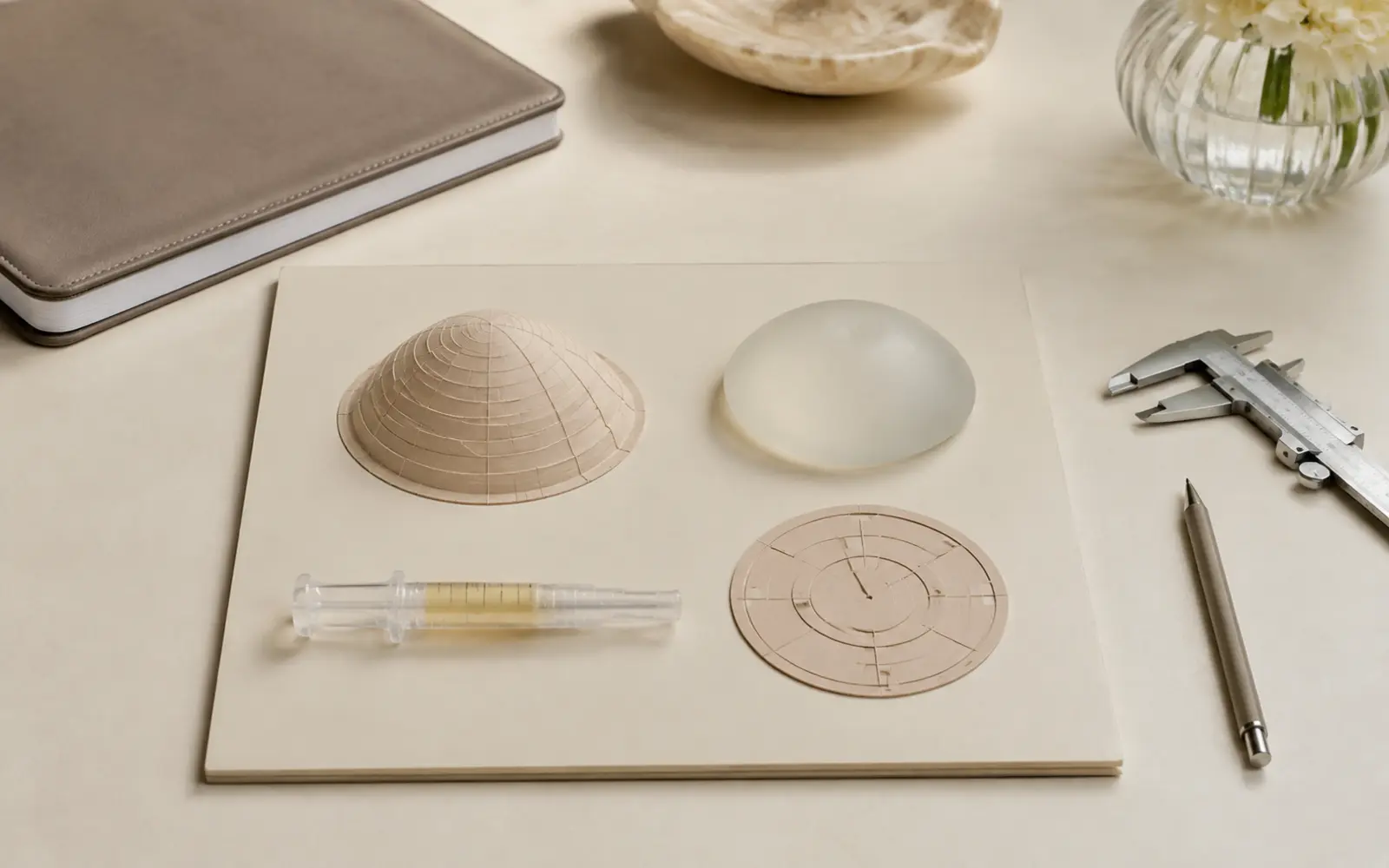
Many mild or moderate patterns can be approached in one stage. Severe constriction, thin tissue coverage, major asymmetry or a large volume goal may make staged treatment more controlled. Tissue safety and predictable healing matter more than a promise of a “one-operation solution”.
| Plan | Possible scar location | Specific follow-up focus |
|---|---|---|
| Base release + implant | Usually in the inframammary fold | Fold position, implant pocket, double contour and symmetry |
| Areolar correction | Around the areola | Tension, scar width and areolar shape |
| Lift or reduction | Periareolar, vertical or inverted-T | Wound healing, nipple blood supply and shape |
| Fat grafting | Small entry points plus donor area | Fat retention, contour and donor-site recovery |
Bleeding, infection, delayed wound healing, altered sensation, asymmetry, contour irregularity, fat necrosis and revision may be discussed with any surgical plan. If implants are used, implant-specific issues include capsular contracture, displacement, rupture and future replacement or removal.
Swelling, tightness and temporary side-to-side differences are common early on. Support garments, showering, sleep position, work, exercise and flying are scheduled according to the operation and personal recovery—not a universal internet timetable.
Pregnancy and weight change can alter breast tissue and skin again. Glandular tissue varies in a tuberous breast, and surgery can also influence breastfeeding. A full milk supply cannot be guaranteed before any breast operation.
Tuberous breasts are generally a developmental breast-shape variation that becomes apparent during puberty. They do not by themselves mean cancer or an acquired disease, but a new lump, discharge, skin tethering or rapid change needs separate medical assessment.
No. Asymmetry may involve volume or level alone. A tuberous shape can combine a constricted base, lower-pole underdevelopment, a high inframammary fold and areolar herniation.
Not always. An implant may add volume, but the constricted base may also need release, lower-pole expansion, fold repositioning or areolar correction.
It may be considered for limited contour differences in selected mild cases. Marked base constriction may require staged grafting or a combination with other techniques.
Scar location depends on the plan. Inframammary, periareolar or vertical patterns may be discussed, and complete disappearance of scars cannot be promised.
The realistic aim is improved balance rather than perfect mirror-image symmetry. The chest wall, tissue quality and healing can differ between sides.
The amount of glandular tissue and the chosen operation may influence breastfeeding. Future pregnancy and feeding plans should be discussed before surgery; a full milk supply cannot be guaranteed.
The stay depends on whether treatment is single-stage or staged, whether an implant or additional reshaping is used, and the early review schedule. A personal timeline is set after examination.
A consultation can assess the base, lower pole, fold, areola and side-to-side differences together.
Op. Dr. Bahadır Çelik: This page was prepared for patient education by the clinic of Op. Dr. Bahadır Çelik, Specialist in Plastic, Reconstructive and Aesthetic Surgery in Antalya. Online information cannot diagnose a breast shape or replace an individual surgical plan; development, tissue quality, imaging needs and suitable options require a medical examination.
This content is for general patient education. Personal suitability, procedure scope, risks and follow-up must be determined after examination.
About us · Contact and appointment · Privacy Policy · Privacy notice